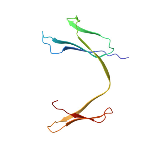
| Link type | Probability | Chain A piercings | Chain B piercings | ||||||||||
|---|---|---|---|---|---|---|---|---|---|---|---|---|---|
| view details |
|
Hopf.2 | 43% | +8B -26B -64B | -36A | ||||||||
| view details |
|
Hopf.2 | 43% | +8B -26B -64B | -36A | ||||||||
| view details |
|
Hopf.2 | 43% | +8B -26B -64B | -36A | ||||||||
| view details |
|
Hopf.2 | 43% | +8B -26B -64B | -36A | ||||||||
| view details |
|
Hopf.2 | 43% | +8B -26B -64B | -36A | ||||||||
| view details |
|
Hopf.2 | 43% | +8B -26B -64B | -36A | ||||||||
| view details |
|
Hopf.2 | 43% | +8B -26B -64B | -36A | ||||||||
| view details |
|
Hopf.2 | 43% | +8B -26B -64B | -36A | ||||||||
Chain A Sequence |
MIQRTPKIQVYSRHPAENGKSNFLNCYVSGFHPSDIEVDLLKNGERIEKVEHSDLSFSKDWSFYLLYYTEFTPTEKDEYACRVNHVTLSQPKIVKWDRDM |
Chain A Sequence |
MIQRTPKIQVYSRHPAENGKSNFLNCYVSGFHPSDIEVDLLKNGERIEKVEHSDLSFSKDWSFYLLYYTEFTPTEKDEYACRVNHVTLSQPKIVKWDR |
| sequence length | 100,98 |
| structure length | 100,98 |
| publication title |
Beta2-microglobulin forms three-dimensional domain-swapped amyloid fibrils with disulfide linkages.
pubmed doi rcsb |
| molecule tags | Protein fibril |
| molecule keywords | Beta-2-microglobulin |
| source organism | Homo sapiens |
| pdb deposition date | 2010-02-04 |
| LinkProt deposition date | 2016-10-29 |
| chain | Pfam Accession Code | Pfam Family Identifier | Pfam Description |
|---|---|---|---|
| AB | PF07654 | C1-set | Immunoglobulin C1-set domain |
| AB | PF07654 | C1-set | Immunoglobulin C1-set domain |
 Image from the rcsb pdb (www.rcsb.org)
#similar chains in the LinkProt database (?% sequence similarity)
...loading similar chains, please wait...
#similar chains, but unlinked
...loading similar chains, please wait...
#similar chains in the pdb database (?% sequence similarity)
...loading similar chains, please wait...